 |
|
|
|
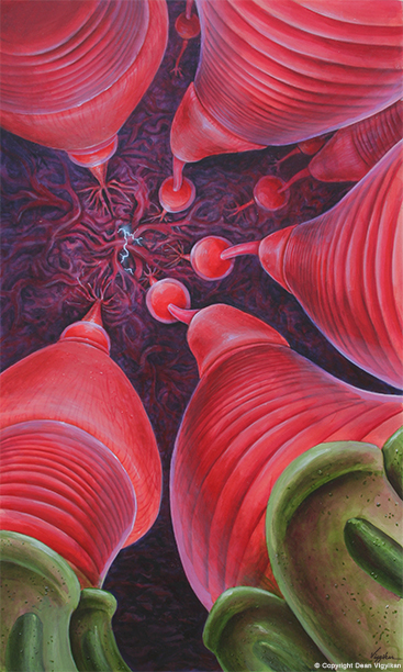
Rods and Cones – Acrylic on Illustration Board
llustrating for the medical and scientific community requires a high degree of accuracy. And there’s also room for imagination. In fact, visual imagination plays a vital role when it comes to determining how to portray objects, concepts, and phenomena which cannot be photographed or viewed with the naked eye. And for some applications, such as entertainment media and advertising, conveying a sense of sheer drama is the name of the game.
This painting is a dramatization and artistic interpretation of the specialized photoreceptor cells, known as rods and cones, within the retina of the eye. The external limiting membrane is omitted in order to allow us a full view of the rods and cones. It’s as though the we were looking up from the pigmented epithelial layer at the very back of the retina and viewing a “ceiling” which represents the outer plexiform layer. The lightning bolt suggests synaptic connections being made between the photoreceptors, stimulated by light, and the dendrites in the outer plexiform layer. It’s amazing to consider that within the retina (which is about .5 mm thick or less) there are yet an additional five structural layers even beyond the outer plexiform layer.